Spontaneously Resolved Lumbar Artery Injury after Blunt Trauma
Article information
Abstract
Major bleeding caused by vascular injuries of the abdominal aorta or its branches after blunt trauma often leads to mortality or major morbidity. We report a case that lumbar artery injury following blunt trauma was spontaneously resolved without any surgical or interventional treatment. Lumbar artery injury after blunt trauma could be treated conservatively without surgical or interventional treatment in a selected case. When an aortic or its branch injury was suspicious, diagnostic angiograms in the setting of interventional treatment may be helpful to decide an appropriate treatment modality.
Introduction
Abdominal vascular injuries may be caused by blunt trauma with rapid deceleration in motor vehicle collisions [1]. Of such vascular injuries, lumbar artery injury (LAI) often leads to mortality and major morbidity due to severity of vascular trauma per se or combined severe trauma [2]. Most studies concerning LAI have been announced as case reports or series. Referring to those studies, LAIs were mostly treated with endovascular procedures such as catheter embolization since surgical intervention often fails to identify bleeding sources, complicating further bleeding [3]. Additionally, more accurate diagnosis of the LAI should be necessary to decide an appropriate treatment modality, because it sometimes mimics abdominal aortic injury or could be even neglected. We reported a case that LAI following blunt trauma was spontaneously resolved without any surgical or interventional treatment.
Case Report
A 63-year old female visited our emergency room for blunt trauma caused by vehicle accident. She complained of pain in the abdomen, the right chest wall and the left forearm. Vital instability was not noted, and the initial blood pressure was 180/100 mmHg on arrival at the emergency room. Focused assessment with sonography for trauma and imaging studies were performed to confirm further concealed trauma. On abdominal computed tomograms, large amount of hematoma adjacent to the infrarenal abdominal aorta with extravasation of contrast material was noted (Fig. 1). Lowering the blood pressure with intravenous beta blockade and preparing blood products for transfusion, she was urgently transferred to the catheterization room to rule out and treat for vascular injury of the abdominal aorta or its branches. On diagnostic angiograms, extravasation of contrast material was not noted, and the abdominal aortic wall seemed to be intact (Fig. 2). We decided to treat her conservatively with serial image follow-up. On the retrospective review of the initial computed tomograms, the LAI was suspected (Fig. 1B). On the 10th hospital day, decrement of the hematoma adjacent to the abdominal aorta was noted without any evidence of further active bleeding on the computed tomographic aortograms (Fig. 3A). She was discharged without any complication and followed up for three months (Fig. 3B).
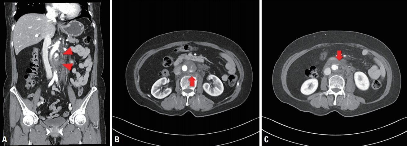
Extravasation of contrast material adjacent to the infrarenal abdominal aorta on abdominal computed tomograms. (A) Arrow heads indicate extravasation of contrast with hematoma on the left side of the infrarenal abdominal aorta. (B) Contrast leakage from lumbar artery was suspected on an axial view (arrow). (C) The inferior mesenteric artery was intact without evidence of contrast leakage on an axial view (arrow).
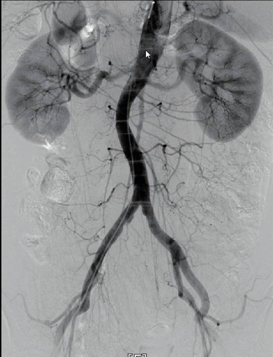
Diagnostic angiograms showed no extravasation of contrast material and intact abdominal aortic wall.
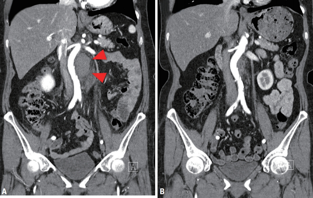
Follow-up computed tomograms. (A) The size of hematoma adjacent to the abdominal aorta decreased without any evidence of further ongoing bleeding on the 10th hospital day (arrow heads). (B) Hematoma adjacent to the abdominal aorta was nearly disappeared three months after blunt trauma.
Discussion
LAI caused by blunt trauma is uncommon and is usually associated with severe abdominal injuries including intraabdominal organ, spinal or pelvic injuries [2]. Isolated LAI without other abdominal injuries is rare. According to prior reports, most of the abdominal vascular injuries by blunt trauma resulted from motor vehicle crashes [1]. Rapid deceleration in motor vehicle collisions may cause two different types of abdominal vascular injuries; avulsion of small branches from major vessels with subsequent hemorrhage and intimal tear with secondary thrombosis of the lumen [1]. This current case seems to be associated with the former mechanism leading to subsequent hemorrhage. Additionally, direct blunt force to the abdomen or back may be the predominant mechanism of abdominal vascular injury, in association with seat-belt use [4,5].
Clinical manifestations of LAI show various spectrum according to its extent. Sometimes it could be resolved spontaneously with conservative treatment just like our case, and sometimes it could lead to more critical conditions necessitating further treatment [6]. It is difficult to decide treatment modality based solely on the initial imaging study. Not only vital status but also consecutive imaging study may be helpful to decide an appropriate treatment option. In this current case, initial computed tomograms showed suspicious ongoing vascular injury of the abdominal aorta or its branches; however, active bleeding was not shown any more on consecutive angiograms. Especially, patient’s vital stability was maintained while staying in the emergency room and that would be noteworthy. However, even stable or asymptomatic patients could be later diagnosed as lumbar artery pseudoaneurysm [6]. Therefore, follow-up imaging studies should be necessary not to miss detection of delayed pseudoaneurysm. In our case, the patient has been followed up for 3 months after hospital discharge and any evidence of lumbar artery pseudoaneurysm was not noted on the latest exams. Optimal duration of imaging follow-ups was not well established; however, imaging studies on the second or third day, 1, 6, and 12 months after the initial study were reasonable referring to the other guidelines concerning aortic diseases [7].
Clinical outcomes of such vascular injury are often devastating. Surgical exploration for LAI is challenging due to difficulty in proximal control and combined injuries [3]. Recently, endovascular interventions were preferred to surgical interventions to improve clinical outcome. Despite such effort, the mortality of abdominal vascular injuries including lumbar artery injury has been reported to be still high and optimal treatment modality, like surgical/endovascular approach, is determined by several factors, like injury pattern, hemodynamic instability, institutional resources, etc [2,4]. Nevertheless, diagnostic angiography and subsequent endovascular treatment for LAI seem to be a good option, especially after stabilization of vital status [8].
Stable LAI by blunt trauma could be treated conservatively without surgical or interventional treatment in a selected case. When an aortic or its branch injury was suspicious, diagnostic angiograms in the setting of interventional treatment may be helpful to decide an appropriate treatment option.