Evaluation of the accuracy of mobile cone-beam computed tomography after spinal instrumentation surgery
Article information

Abstract
Purpose
Pedicle screw fixation provides 3-column stabilization, multidimensional control, and a higher rate of interbody fusion. Although computed tomography (CT) is recommended for the postoperative assessment of pedicle screw fixation, its use is limited due to the radiation exposure dose. The purpose of this preliminary retrospective study was to assess the clinical usefulness of low-dose mobile cone-beam CT (CBCT) for the postoperative evaluation of pedicle screw fixation.
Methods
The author retrospectively reviewed postoperative mobile CBCT images of 15 patients who underwent posterior pedicle screw fixation for spinal disease from November 2019 to April 2020. Pedicle screw placement was assessed for breaches of the bony structures. The breaches were graded based on the Heary classification.
Results
The patients included 11 men and four women, and their mean age was 66±12 years. Of the 122 pedicle screws, 34 (27.9%) were inserted in the thoracic segment (from T7 to T12), 82 (67.2%) in the lumbar segment (from L1 to L5), and six (4.9%) in the first sacral segment. Although there were metal-related artifacts, the image of the screw position (according to Heary classification) after surgery could be assessed using mobile CBCT at all levels (T7–S1).
Conclusions
Mobile CBCT was accurate in determining the location and integrity of the pedicle screw and identifying the surrounding bony structures. In the postoperative setting, mobile CBCT can be used as a primary modality for assessing the accuracy of pedicle screw fixation and detecting postoperative complications.
INTRODUCTION
Pedicle screw fixation provides three-column stabilization, multidimensional control, and a greater rate of interbody fusion [1]. It is ideal for the screw to be fully contained within the pedicle without breaching it. Inaccurate pedicle screw fixation with a breach can lead to serious complications such as nervous or connective tissue injuries and spinal instability [2]. Although computed tomography (CT) is considered to be the most useful imaging modality for the postoperative assessment of pedicle screw fixation, its use is limited due to the radiation exposure dose. Although multi-detector CT (MDCT) is currently used in most hospitals, its use to evaluate surgical outcomes and prognoses during follow-up increases the radiation exposure dose received by patients in proportion to the number of tests. Cone-beam CT (CBCT), in contrast, has a significantly lower radiation exposure dose than medical MDCT because it can obtain a high-definition image with a single scan using a flat panel detector. The purpose of this preliminary retrospective study was to assess the clinical usefulness of postoperative evaluation of pedicle screw fixation using low-dose mobile CBCT.
METHODS
This study was approved by the Institutional Ethical Committee of the Wonkwang University Hospital and was conducted in compliance with the institution’s requirements (No. 202002021).
The authors retrospectively reviewed the postoperative mobile CBCT images of 15 patients who underwent posterior pedicle screw fixation for spinal disease between November 2019 and April 2020. All operations were performed by one of two neurosurgeons. The inclusion criteria were patients with an adult spinal disorder over the age of 19 years, not falling under the category of vulnerable subjects, the willingness to participate in a clinical trial based on voluntary consent, and the ability to undergo CT. The exclusion criteria were unstable vital signs, pregnancy, having a spine containing a substance that could affect image acquisition, and having been judged as unsuitable for participation in clinical trials due to other reasons.
Mobile CBCT scanners and imaging interpretation
The use of two axis detectors with a larger area and a cone-shaped X-ray beam with a large aperture angle (widely divergent X-ray beam) is the specific feature of CBCT scanners. CBCT makes it possible to obtain a three-dimensional image with high spatial resolution in the course of a single rotation of the emitter and detector without moving the patient through the gantry. The radiation dose received in the course of CBCT is much lower than that received during MDCT [3,4]. CBCT has been widely used for the diagnosis of diseases and injuries of the maxillofacial, ear, nose, and throat regions for about 20 years. Due to its advantages, CBCT also holds considerable promise for examining orthopedic patients. In recent years, several manufacturing companies have developed specialized CBCT scanners for extremity imaging, which, among other advantages, allow carrying out studies under loading with patients in a vertical position [3,5]. Mobile CBCT scans were obtained using MX-CBT1240 (Phion 2.0; NanoFocusRay, Iksan, Korea) (Fig. 1). The components of the MX-CBT1240 system included a high-frequency generator, a rotating anode X-ray tube, and an amorphous silicon thin-film transistor flat panel detector. The system had the following specifications: scan time, 7–13 seconds; bore size, 650 mm; single scan, 360° rotation; field of view, transaxial 260 mm and length 165 mm; reconstruction time, <41 seconds; AC power, 200–230 V/13 A, 50/60 Hz; weight, 400 kg. The typical scanning parameters were 110 kV, 20 mA, one pitch, a slice thickness of 3 mm, and a rotation time of 20 milliseconds.
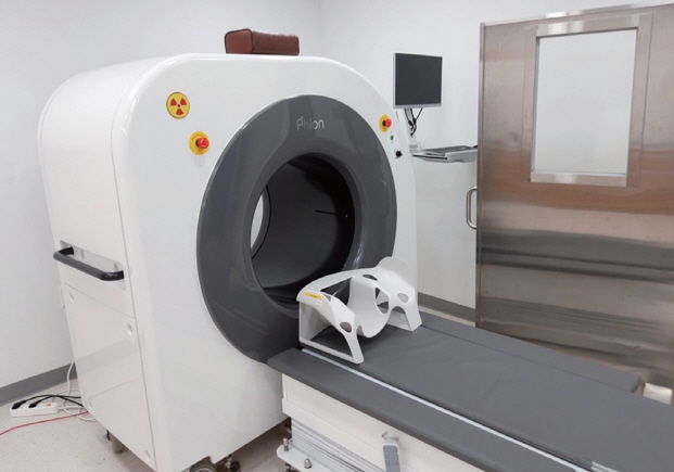
Mobile cone-beam computed tomography using MX-CBT1240 (Phion 2.0; NanoFocusRay, Iksan, Korea).
Surgical technique
The surgical procedures involved open standard posterior transpedicular screw fixation with the patient in the prone position. The surgeon checked the surgical level using the C-arm, and the pedicle screws were inserted based on the anatomic landmarks under the C-arm. After awakening the patient from anesthesia after surgery, it was confirmed that the patient’s vital signs stabilized in the recovery room. The patient was moved to a mobile CT room located next to the recovery room and postoperative mobile CBCT was taken. Mobile CBCT was performed to assess the screw positions and surrounding bony structure after surgery.
Accuracy of pedicle screw insertion
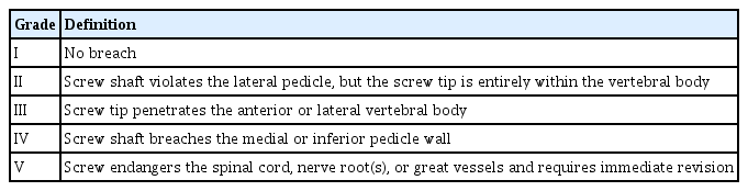
The accuracy of pedicle screw placement was assessed based on the breach of bony structures. Breaches were graded according to the Heary classification (Table 1) [6]. The Heary classification includes five grades: grade I refers to a screw completely contained within the pedicle; grade II refers to an in-out-in screw with a lateral breach, with a screw tip completely contained within the vertebral body; grade III refers to a pedicle screw with a tip that penetrates the anterior or lateral vertebral body; grade IV refers to medial and inferior pedicle breaches; and grade V refers to a screw that endangers neural or vascular structures and requires immediate repositioning. Three patients with grade III had no specific clinical symptoms and did not require additional treatment.
RESULTS
Demographic characteristics
A total of 15 patients who underwent posterior transpedicular screw fixation for spinal trauma or diseases were included in this preliminary study. The demographic characteristics of the patients are summarized in Table 2. The patients included 11 men (73.3%) and four women (26.7%), and their mean age was 66±12 years (range, 41–74 years). Their mean weight and height were 71±10 kg and 168±15 cm, respectively. The diagnosis was trauma in ten patients (66.7%) and degenerative disease in five (33.3%). The mean surgical duration was 199±42 minutes (range, 121–368 minutes). Of the 122 pedicle screws, 34 (27.9%) were inserted in the thoracic segment (from T7 to T12), 82 (67.2%) in the lumbar segment (from L1 to L5), and six (4.9%) in the first sacral segment.
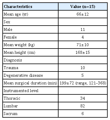
Demographic characteristics of patients
Breaches according to the Heary classification
The number of pedicle screws at each vertebral level and the results of the screw placement assessment using postoperative mobile CBCT imaging are shown in Table 3. The grading of the screws based on the Heary classification was as follows: 106 of the screws (86.9%) were placed within the pedicle without any breach (grade I); 13 (10.7%) were in-out-in screws with a lateral breach, and the screw tip was inside the vertebral body (grade II); three (2.5%) had an anterior or lateral breach (grade III); none had a medial breach (grade IV); and none had a breach that required immediate revision (grade V).

Number of pedicle screws at each vertebral level and the frequency of breaches on postoperative mobile cone-beam computed tomography images based on the Heary classification
Imagery interpretation using mobile CBCT
Although there were some metal-related artifacts, the images of the screw position (according to Heary classification) after surgery using mobile CBCT at all levels (T7–S1) were readable.
Illustrative case
A 46-year-old male patient reported to the hospital with low back pain after falling from a height of 5 m. He had undergone posterior lumbar interbody fusion (PLIF) with transpedicular screw fixation at L3–L4 for spinal stenosis at another hospital 5 years earlier. A physical examination revealed severe direct tenderness at the thoracolumbar junction. On neurological examination, he had numbness and paresthesia in both legs. Lumbar CT revealed an acute burst fracture of the L1 body with mild depression and previous PLIF at L3–L4 (Fig. 2A, B). Lumbar magnetic resonance imaging (MRI) revealed an acute burst fracture with a partial tear of the posterolateral ligament complex at L1 and an epidural hematoma extending from T11 to L2/3 (Fig. 2C, D). Preoperative evaluations, including electrocardiography, cardiac sonography, pulmonary function tests, and laboratory tests, revealed no abnormalities. He underwent posterior transpedicular screw fixation at T12, L1, and L2. Postoperative mobile CBCT revealed that the screw shaft violated the lateral pedicle, and the screw tip was entirely within the vertebral body of the right L1. However, the screw shaft and the tip of the left L1 were entirely within the pedicle and vertebral body without breach (Fig. 2E–G). The postoperative course was uneventful, and the patient was discharged without any neurological defects 14 days after surgery.

(A) Lumbar axial and (B) sagittal computed tomography showing an acute burst fracture of the L1 body with mild depression and a previous posterior lumbar interbody fusion with pedicular screw fixation at L3–L4. (C) Lumbar axial and (D) sagittal magnetic resonance images showing an acute burst fracture with a partial tear of the posterolateral ligament complex at L1 and an epidural hematoma extending from T11 to L2/3. (E) Postoperative axial, (F) coronal, and (G) sagittal mobile cone-beam computed tomography showing violation of the lateral pedicle by the screw shaft and the screw tip entirely within the vertebral body of the right L1. The screw shaft and the tip of the left L1 are entirely within the pedicle and vertebral body without a breach. The patient provided written informed consent for the publication of the research details and clinical images
DISCUSSION
Pedicle screw fixation, which was first described by Boucher in the 1950s and further investigated by Roy-Camille later in the 1960s and 1970s, is extensively used for spinal fixation and stabilization in several spinal diseases, including spinal fracture, deformities, spondylitis, and degenerative diseases [2,7,8]. The screw should be inserted within the central part of the pedicle, and it should enter the vertebral body parallel to the endplate without breaching the cortex, although sacral screws can penetrate the anterior cortex for short distances [9]. Misplacement of the pedicle screw can lead to nerve root injury, pedicle fracture, dural tear injury with cerebrospinal fluid leakage, vascular injury, visceral injury from screw overpenetration, and facet joint violation [1,2]. All of these complications lead to spinal pain or disability in patients and increase the need for reoperation and related healthcare costs [2]. The rate of screw malpositioning in this study was 13.1%; although the reported rate of screw malpositioning varies considerably in the literature, it may be as high as 40% [10]. This variation may be associated with several factors, such as the surgeon’s experience, techniques, and differences in imaging devices. Several intraoperative techniques have been developed in recent years to improve the accuracy and safety of pedicle screw placement. However, the versatility of the shape and dimensions of the spine makes it difficult to place the pedicle screws accurately in some cases [11].
Most spine surgeons may still use the so-called conventional freehand technique, which tends to be based on the use of image intensifiers in at least one plane [11]. The conventional insertion of the pedicle screw is performed by identifying the entry point and directly palpating the pedicle wall using a probe [2]. Radiological imaging equipment can be used to increase the accuracy of pedicle screw placement. Among the various types of intraoperative imaging equipment, the C-arm is most commonly used because it is easy to use, familiar, and relatively inexpensive. Because the C-arm is used for two-dimensional fluoroscopy and it has a low resolution, the accuracy of screw insertion may be poor in complex anatomical areas. Although the fluoroscopic C-arm may be acceptable for pedicle screw fixation, more advanced equipment such as CT is recommended for higher accuracy [11,12]. However, CT-guided pedicle screw insertions for spinal disease have several disadvantages, including the radiation exposure of patients, surgeons, and operating room staff, which is significantly higher than that of intraoperative C-arm fluoroscopy. The surgery duration and the risk of infection can also increase. A follow-up CT examination to evaluate the position of the pedicle screw after surgery can also increase patients’ radiation exposure. CT is therefore considered more useful for postoperative evaluation of the insertion of the screw and follow-up observations after surgery rather than for use during surgery.
The frequency of spinal surgery has steadily increased over the past decades due to innovations in surgical techniques and devices. X-rays are a primary imaging modality used for postoperative evaluation and regular long-term follow-up after spinal pedicle screw fixation.
Although X-ray examinations for spine surgery have some limitations, they can often be used to check the location of instruments and the degree of bone fusion and diagnose certain complications such as fractures and deformities [13]. Comparing the subsequent X-ray findings with the immediate postoperative radiographic findings is important for detecting changes in the inserted spine instruments and adjacent bony structures. Although X-ray examinations and CT are commonly used to evaluate pedicle screw placement postoperatively, their accuracy has been debated. Farber et al. [14] used postoperative radiographs and CT to evaluate the placement of 74 pedicle screws in 16 consecutive patients undergoing lumbar pedicle screw fixation. In their series, fewer screws were clearly within the pedicle on CT than on radiograph, and CT showed 10 times as many screws violating the medial cortex as did radiographs. They showed that conventional radiographs alone may not accurately reveal pedicle screw placement. Laine et al. [15] performed a prospective study of the accuracy of pedicle screw placement in 30 low back operations. The total number of screws was 152. CT imaging diagnosed a total of 32 misplaced pedicle screws (21%), whereas conventional radiographs diagnosed only four of these misplaced placements (3%). They concluded that conventional radiographs give a false impression of accuracy and safety in pedicular screw placement. Learch et al. [16] reported that 63% of screw placements were correctly identified as in or out of the pedicle using conventional radiographs, whereas CT improved the accuracy to 87%. Although ultrasonography can be used to detect superficial fluid collection, hematoma, and abscess, it has several limitations for assessing the surgical results of pedicle screw fixation [9]. MRI also has limitations in evaluating the postoperative state of pedicle screw fixation due to artifacts from metallic implants. CT is appropriate for postoperatively assessing pedicle screw fixation and detecting postoperative complications. CT also provides a very detailed description of the bone structure and uses a multi-plane reconstruction function that improves the location and alignment of implants and facilitates the evaluation of bone fusion through a high spatial resolution isotropic dataset [9]. When X-ray beams pass through a metallic screw, photon starvation, beam hardening, and beam scattering occur, and distinct dark and bright bands may appear as artifacts in a CT image. These artifacts limit the visibility of the screws and surrounding bones [17]. To reduce the metal artifacts and improve image quality, an optimized CT protocol and advanced artifact reduction technology are required. For CT to be a more useful device for postoperative evaluation of spine patients, the amount of irradiation should be small, the image should be acquired quickly, and the movement of the equipment should be convenient. The radiation exposure dose of mobile CBCT used in this study was a mean volume computed tomography dose index (CTDIvol) of 2.9 mGy. The radiation exposure dose of MDCT used in spine imaging is a CTDIvol of about 10 mGy. Although this difference in the diagnostic radiation dose does not directly affect the human body, the accumulation of radiation doses from repeated exposure can cause serious health problems, including cancer [18]. Brenner and Hall [18] reported direct evidence from epidemiological studies that the organ doses corresponding to common CT studies (two or three scans) resulted in an increased risk of cancer, which was reasonably convincing for adults and very convincing for children. Economic considerations should also be kept in mind. In Korea, medical insurance currently covers mobile CBCT for limb joints, but not for spinal images. In addition, using mobile CBCT in a recovery room with radiation shielding facilities would have the advantage of enabling rapid identification and treatment of screw malposition. We believe that these studies will provide a basis for discussing insurance coverage of mobile CBCT spinal imaging after sufficient verification of its clinical usefulness and safety. The most important prerequisite for CT to be a more useful device for postoperative spinal evaluation is to obtain a high-quality image that can accurately confirm the screw position in the spine through low-dose CT.
Study limitations
There are several limitations of this study. First, the number of cases is small for judging the accuracy of pedicle screw placement. Additional research is required to compare and evaluate a larger number of pedicle screws. Second, as a postoperative assessment of pedicle screw fixation, the accuracy of mobile CBCT was not compared with that of other imaging devices such as conventional radiographs, MDCT, and MRI. Third, there are several limitations in evaluating the usefulness of CBCT because there no comparison was made between MDCT and mobile CBCT in terms of the time required for the CT scan, radiation exposure, and convenience.
In conclusions, mobile CBCT was accurate for determining the location and integrity of the pedicle screw and identifying the surrounding bony structures. The authors could obtain high-quality images within a short scan duration with a low dose using mobile CBCT. In the postoperative setting, mobile CBCT can be used as a primary modality for assessing the accuracy of pedicle screw fixation and detecting postoperative complications. Further research is warranted to evaluate the usefulness of mobile CBCT compared to MDCT.
Notes
Ethical statements
The study was approved by the Institutional Ethical Committee of the Wonkwang University Hospital and was conducted in compliance with the institution’s requirements (No. 202002021). Informed consent was obtained from all individual participants included in this study.
Conflicts of interest
Kwon-Ha Yoon is the CEO of NanoFocusRay, Iksan, Korea. The authors have no other conflicts of interest to declare.
Funding
None.
Author contributions
Conceptualization: KSE, KHY; Data curation: ESP, JTP; Formal analysis: KSE, ESP; Funding acquisition: JTP, KHY; Investigation: KSE, DWK; Methodology: KSE, DWK; Project administration: JTP, KHY.
All authors read and approved the final copy of the manuscript.