
Articles
- Page Path
- HOME > J Trauma Inj > Volume 37(1); 2024 > Article
-
Case Report
Misinterpretation of a skin fold artifact as pneumothorax on the chest x-ray of a trauma patient in Korea: a case report -
Yoojin Park, MD1
 , Eun Young Kim, MD1, Byungchul Yu, MD2, Kunwoo Kim, MD3
 -
Journal of Trauma and Injury 2024;37(1):86-88.
DOI: https://doi.org/10.20408/jti.2023.0050
Published online: February 23, 2024
- 495 Views
- 6 Download
1Department of Radiology, Gachon University Gil Medical Center, Gachon University College of Medicine, Incheon, Korea
2Department of Trauma Surgery, Gachon University Gil Medical Center, Gachon University College of Medicine, Incheon, Korea
3Department of Thoracic and Cardiovascular Surgery, Gachon University Gil Medical Center, Gachon University College of Medicine, Incheon, Korea
- Correspondence to Eun Young Kim, MD Department of Radiology, Gachon University Gil Medical Center, Gachon University College of Medicine, 21 Namdong-daero 774beon-gil, Namdong-gu, Incheon 21565, Korea Tel: +82-32-460-3060 Email: oneshot0229@gilhospital.com
© 2024 The Korean Society of Traumatology
This is an Open Access article distributed under the terms of the Creative Commons Attribution Non-Commercial License (http://creativecommons.org/licenses/by-nc/4.0/) which permits unrestricted non-commercial use, distribution, and reproduction in any medium, provided the original work is properly cited.
ABSTRACT
- Misinterpreting radiographic findings can lead to unnecessary interventions and potential patient harm. The urgency required when responding to the compromised health of trauma patients can increase the likelihood of misinterpreting chest x-rays in critical situations. We present the case report of a trauma patient whose skin fold artifacts were mistaken for pneumothorax on a follow-up chest x-ray, resulting in unnecessary chest tube insertion. We hope to help others differentiate between skin folds and pneumothorax on the chest x-rays of trauma patients by considering factors such as location, shape, sharpness, and vascular markings.
- Chest x-ray (CXR) is the most readily available and common imaging modality for trauma patients, providing information on the patient's overall thoracic health and helping to identify thoracic injuries [1]. However, the interpretation of CXRs can be challenging, particularly when adequate image quality is difficult to obtain in a seriously injured patient [2]. The likelihood of image artifact and misinterpretation of the CXR increases when the patient has multiple monitoring and resuscitation lines and devices in the chest [3]. According to previous reports, approximately 10% of errors occurred during interpretation of the CXR, as the interpretation of CXRs is subject to human error and depends on reader expertise [4].
- A skin fold artifact is one of the commonly encountered artifacts on CXR that can mimic pneumothorax [5]. Failure to differentiate between true pneumothorax and a skin fold artifact can lead to unnecessary interventions and complications, such as iatrogenic bleeding, prolonged hospital stays, and increased healthcare costs [6].
INTRODUCTION
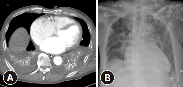
- A 72-year-old man presented to the emergency department with injuries from a fall. The initial assessment showed multiple bilateral rib and spine fractures as well as pelvic bone fractures. The initial chest computed tomography and CXR (Fig. 1) showed bilateral hemothorax, so chest tubes were inserted (not shown here). After 6 days of hospitalization, the right chest tube was removed due to decreased hemothorax (Fig. 2A). However, after 8 days of hospitalization, acute pneumothorax was suspected (Fig. 2B) on the follow-up CXR, prompting bedside chest tube reinsertion (Fig. 2C). As there was no evidence of air drainage from the chest tube and a repeat CXR the next day showed no pneumothorax, interdepartment consultation was requested with radiology. The thoracic radiologists confirmed that it was a skin fold and not an actual pneumothorax, leading to removal of the chest tube. The follow-up CXR (Fig. 2D) again showed multiple skin folds from a different view.
- Ethics statement
- This study was approved by the Institutional Review Board of Gachon University College of Medicine (No. GAIRB2023-216), which waived the requirement for informed consent.
CASE REPORT
- Chest radiography is a common diagnostic test used to evaluate chest pathologies such as pneumothorax, hemothorax, pneumonia, and lung injury in injured patients since it allows easy and fast assessment [7,8]. However, normal structures such as nipples and skin folds and clothing items like buttons or attached electrocardiography leads can be misinterpreted as abnormal findings on CXR [9–11]. Skin fold artifacts are commonly encountered on CXRs, especially in patients with obesity or those unable to cooperate with optimal positioning during the examination [12,13]. These artifacts can mimic various pathological conditions, including pneumothorax, leading to potential misdiagnosis and unnecessary interventions. Therefore, clinicians must be able to recognize these artifacts and pay special attention when interpreting the CXRs of traumatized patients.
- Some tips for differentiating pneumothorax from a skin fold on CXR include the following [14,15]:
- 1. Location: Pneumothorax typically appears along the lung periphery, whereas a skin fold commonly appears to continue beyond the chest wall.
- 2. Shape and sharpness: The linear shadow of pneumothorax tends to be thin, sharp, and well-defined. A skin fold usually appears wider and ill-defined towards the medial side, while appearing sharp towards the lateral side.
- 3. Vascular markings: The distal portion of the linear shadow of pneumothorax often lacks visible pulmonary vessels, whereas the distal portion of a skin fold may still demonstrate pulmonary vessel markings.
- In situations where pneumothorax and skin fold can be easily confused, clinicians should consider these radiological tips when making decisions, while also taking into account the clinical context and other relevant factors.
DISCUSSION
-
Conflicts of interest
The authors have no conflicts of interest to declare.
-
Funding
This study was supported by a grant from Gachon University Gil Medical Center (No. FRD2021-11).
-
Author contributions
Conceptualization: EYK; Funding acquisition: EYK; Methodology: YP; Visualization: YP; Writing–original draft: YP; Writing–review & editing: all authors. All authors read and approved the final manuscript.
-
Data availability
Data sharing is not applicable as no new data were created or analyzed in this study.
ARTICLE INFORMATION

- 1. Lewis BT, Herr KD, Hamlin SA, et al. Imaging manifestations of chest trauma. Radiographics 2021;41:1321–34. ArticlePubMedPMCPDF
- 2. Mathew RP, Alexander T, Patel V, Low G. Chest radiographs of cardiac devices (part 1): lines, tubes, non-cardiac medical devices and materials. SA J Radiol 2019;23:1729. ArticlePubMedPMC
- 3. Baratella E, Marrocchio C, Bozzato AM, Roman-Pognuz E, Cova MA. Chest x-ray in intensive care unit patients: what there is to know about thoracic devices. Diagn Interv Radiol 2021;27:633–8. ArticlePubMed
- 4. Gefter WB, Post BA, Hatabu H. Commonly missed findings on chest radiographs: causes and consequences. Chest 2023;163:650–61. ArticlePubMedPMC
- 5. Kishimoto K, Watari T, Tokuda Y. Pseudo-pneumothorax: skin fold is an excellent imitator. BMJ Case Rep 2018;2018:bcr2018226360. ArticlePubMedPMC
- 6. Niazi AK, Minko P, Nahrstedt CJ, et al. A case of pseudo-pneumothorax with complications. Cureus 2018;10:e3263PubMedPMC
- 7. Zarogoulidis P, Kioumis I, Pitsiou G, et al. Pneumothorax: from definition to diagnosis and treatment. J Thorac Dis 2014;6(Suppl 4):S372–6. ArticlePubMed
- 8. Robin ED, Burke CM. Routine chest x-ray examinations. Chest 1986;90:258–62. ArticlePubMed
- 9. Petersen GW, Baier H. Incidence of pulmonary barotrauma in a medical ICU. Crit Care Med 1983;11:67–9. ArticlePubMed
- 10. Chiles C, Ravin CE. Radiographic recognition of pneumothorax in the intensive care unit. Crit Care Med 1986;14:677–80. ArticlePubMed
- 11. Waite S, Scott J, Gale B, Fuchs T, Kolla S, Reede D. Interpretive error in radiology. AJR Am J Roentgenol 2017;208:739–49. ArticlePubMed
- 12. Uppot RN, Sahani DV, Hahn PF, Gervais D, Mueller PR. Impact of obesity on medical imaging and image-guided intervention. AJR Am J Roentgenol 2007;188:433–40. ArticlePubMed
- 13. Eisenhuber E, Schaefer-Prokop CM, Prosch H, Schima W. Bedside chest radiography. Respir Care 2012;57:427–43. ArticlePubMed
- 14. Kattea MO, Lababede O. Differentiating pneumothorax from the common radiographic skinfold artifact. Ann Am Thorac Soc 2015;12:928–31. ArticlePubMed
- 15. Bruno MA, Walker EA, Abujudeh HH. Understanding and confronting our mistakes: the epidemiology of error in radiology and strategies for error reduction. Radiographics 2015;35:1668–76. ArticlePubMed
 PubReader
 ePub Link
 Cite
Cite


